
Punched-Out Papillae at 5pm
Jul 21, 2026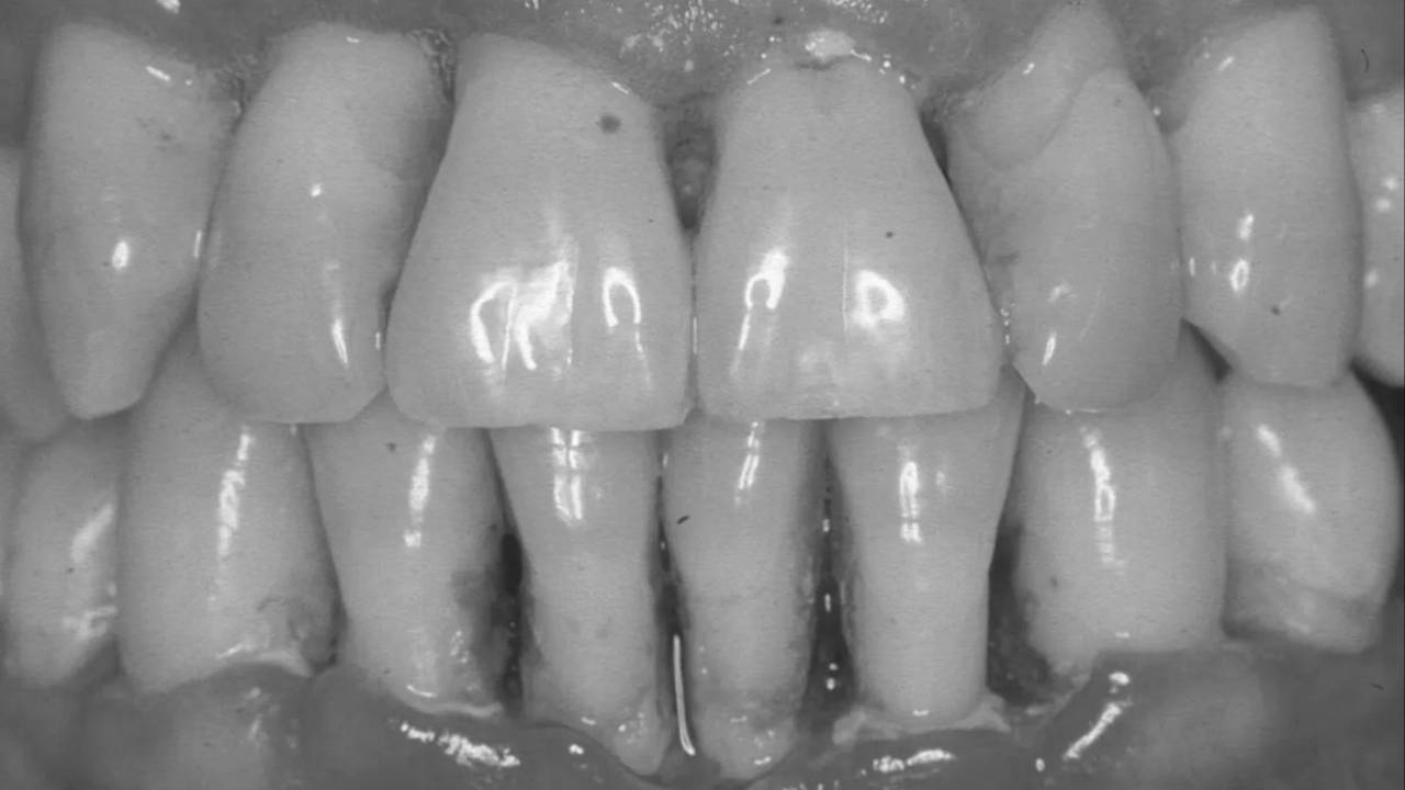
Author: Gum Specialist Dr Reena
It’s 5pm on a Thursday. A patient calls - pain, bleeding gums, a smell they can’t quite describe, and a mouth that looks nothing like it did a week ago. You’ve seen gum disease before. You haven’t seen this. Acute periodontal presentations are uncomfortable precisely because they sit at the intersection of the familiar and the alarming - conditions that look like things you know but behave in ways that catch you off guard if you’re not prepared. The good news is that preparation is straightforward. Here is what you need to know.
The Abscess You Need to Distinguish First
Not all periodontal abscesses are the same - and the first job is making sure you’re looking at the right one. A gingival abscess is a localised purulent infection of the marginal gingiva or interdental papilla, almost always associated with a foreign body impacted into previously healthy tissue. The nail-biter with a swollen papilla. The patient who caught their gum with a toothpick. Drain it, irrigate it, remove the cause. It resolves.
A periodontal abscess is a different animal. Defined as a localised accumulation of pus within the gingival wall of a periodontal pocket, it sits third on the list of the most prevalent infections requiring emergency treatment in UK general practice - behind dento-alveolar abscesses and pericoronitis, but present in 6-7% of patients treated in a single month (Lewis et al., 1989). The presentation is characteristic: an ovoid elevation along the lateral aspect of the root, tenderness on palpation, a deep pocket with bleeding and suppuration on probing, increased mobility, and a patient who reports the tooth feeling “high.” The tooth will be vital on sensibility testing - this is the critical differential from a perio-endo lesion.
Management: establish drainage through the pocket or via external incision, debride the pocket thoroughly, and resist the instinct to prescribe antibiotics. Systemic antimicrobials are only warranted when there is clear evidence of spreading infection - fever, lymphadenopathy, systemic involvement. A localised abscess is a local problem requiring local treatment. The antibiotic is not a substitute for the drainage.
Then there’s the perio-endo lesion - the one most likely to trip you up. The abscess associated with a deep pocket on a non-vital tooth. Sensibility testing is the key: negative response points toward a primary endodontic origin. The communication between pulp and periodontium can run in either direction - pulpal disease expressed through the PDL, or periodontal disease communicating to the pulp apically. Vertical root fracture can present identically, and in a heavily restored tooth with a localised narrow deep pocket, fracture should be high on the differential. A primary endodontic lesion often responds well to root canal therapy alone. A true combined lesion carries a poor prognosis — and honesty with the patient about that prognosis is part of the management.
The One That Looks Like the End of the World
Necrotising periodontal disease is uncommon enough to be unfamiliar and dramatic enough to be immediately alarming. It is also, once you know what you’re looking at, completely unmistakable. Punched-out necrotic papillae. Spontaneous bleeding. A pseudomembrane over the necrotic tissue that, when removed, leaves raw exposed connective tissue beneath. Halitosis that is genuinely unlike anything else. Pain that worsens with eating and oral hygiene. The patient is not overreacting. This is the most severe inflammatory lesion associated with the oral biofilm.
Necrotising gingivitis (NG) affects the gingival tissue only. Necrotising periodontitis (NP) extends into the periodontal ligament and alveolar bone - the necrosis creating interproximal craters that, as they spread and merge, produce an extensive zone of destruction. These may be different stages of the same disease process rather than distinct conditions (Horning & Cohen, 1995).
The predisposing factors tell you as much as the clinical picture. Psychological stress - often acute, recent, significant. Heavy smoking. Poor nutrition. Sleep deprivation. In patients where none of these obvious triggers are present, particularly in a young adult, necrotising disease should prompt consideration of undiagnosed HIV or haematological malignancy. The periodontal presentation may be the first clinical signal. This is not a reason to alarm the patient in the chair - it is a reason to ask the right questions and refer appropriately.
Management: gentle superficial debridement, ultrasonic instruments preferred over hand instruments on ulcerated tissue, getting progressively deeper as the patient’s tolerance improves over 2-4 days. Chlorhexidine 0.2% twice daily. If there is no improvement following debridement, metronidazole 400mg three times daily for five days - the first choice given its action against the strict anaerobes driving the destruction. Adequate analgesia. Nutrition. Address the risk factors, because this is a condition that recurs if the underlying triggers persist.
The 5pm Call About the Wisdom Tooth
Pericoronitis is the most predictable of the acute perio presentations - and still the most mismanaged. Inflammation of the soft tissue surrounding a partially erupted tooth, almost always the lower third molar, where the overlying operculum becomes a harbour for debris, bacteria and trauma from the opposing tooth. Mild cases: localised swelling, tenderness, bad taste. Severe cases: trismus, extraoral swelling, lymphadenopathy, fever, radiating pain to the ear. These are not the same presentation and they do not warrant the same management.
Acute management: irrigate beneath the operculum, remove debris, consider occlusal adjustment on the opposing tooth if it’s traumatising the flap. Antibiotics only if there are systemic signs or evidence of spreading infection — not as a first-line response to a localised swelling. The patient who presents with trismus and facial swelling needs urgent assessment to exclude early space infection. That clinical judgement - pericoronitis vs something more serious - matters.
Long-term management is equally important. Evidence supports that a first episode of pericoronitis, unless particularly severe, is not in itself an indication for surgery (NICE). Second or subsequent episodes are. Have that conversation clearly and early, because the patient who presents for the third time in a year expecting the same antibiotic prescription needs a different plan.
The Things That Aren’t What They Look Like
Acute herpetic gingivostomatitis - the most common viral infection of the oral mucosa - is most frequently seen in children aged two to five but can present at any age. Multiple small ulcers with elevated margins dispersed across both attached and unattached mucosal surfaces, accompanied by systemic features: lymphadenopathy, fever, malaise. The key differential from necrotising gingivitis: the ulcers in HSV affect the entire oral mucosa, not just the interdental papillae; the patient is typically feverish; and the condition lasts 7-10 days and self-resolves. Management is supportive - adequate hydration, analgesia sufficient to allow eating and drinking, and in immunocompromised patients, urgent referral for intravenous antiviral therapy.
Physical, chemical and thermal injuries round out the acute picture and are underappreciated as a category. The overzealous horizontal brusher with an erosion at the gingival margin. The patient who applied bleaching gel with a poorly fitting tray and burned their mucosa. The hot pizza injury that desquamated the palate. These are not biofilm-driven lesions - but accurate diagnosis matters, because the treatment is removal of the cause, not periodontal therapy. The lesion that doesn’t fit the pattern, that has no clear initiating factor, that persists despite the obvious management - that needs oral medicine input, not a reassuring review appointment.
The Fracture You Almost Missed
A subgingival root fracture is the acute presentation most likely to be misdiagnosed - and the consequences of missing it are significant. A localised deep narrow pocket on a tooth that may be minimally restored. Pain on biting, or characteristically on release of biting load. Tenderness to percussion. The radiograph may show nothing. The pocket tells you everything. Magnification, good illumination, a Tooth Slooth device to load individual cusps - these are your diagnostic tools. A periodontal flap may be necessary to confirm the diagnosis and visualise the apical extent of the fracture. In many cases, the tooth is unsavable. Knowing that early, and communicating it clearly, is better than multiple management attempts followed by an inevitable extraction.
The Rule That Applies to All of Them
Diagnose before you prescribe. Every acute periodontal presentation has a cause - a pocket, a foreign body, an operculum, a virus, a fracture, a bleaching agent. Treatment directed at the cause resolves the problem. Antibiotics prescribed instead of diagnosis delay it and contribute to resistance. Analgesia should be adequate - the patient in acute periodontal pain needs relief, not reassurance. And when the clinical picture doesn’t fit, when the presentation is unusually severe, unusually rapid, or associated with systemic features that don’t add up - that is the moment to refer, not the moment to manage conservatively and review in two weeks.
Emergencies clarify. The acute presentation strips away the routine and demands something that elective dentistry rarely requires - immediate, clear-headed decision-making under pressure, with a patient in pain who needs you to know what you’re looking at. The clinician who handles a 5pm periodontal emergency well demonstrates something no elective procedure can: diagnosis first, management second, referral when the situation requires it. That combination - knowledge, judgement, and the self-awareness to know your limits - is exactly what patients need. And it is worth being prepared for.
